Česky
Česky


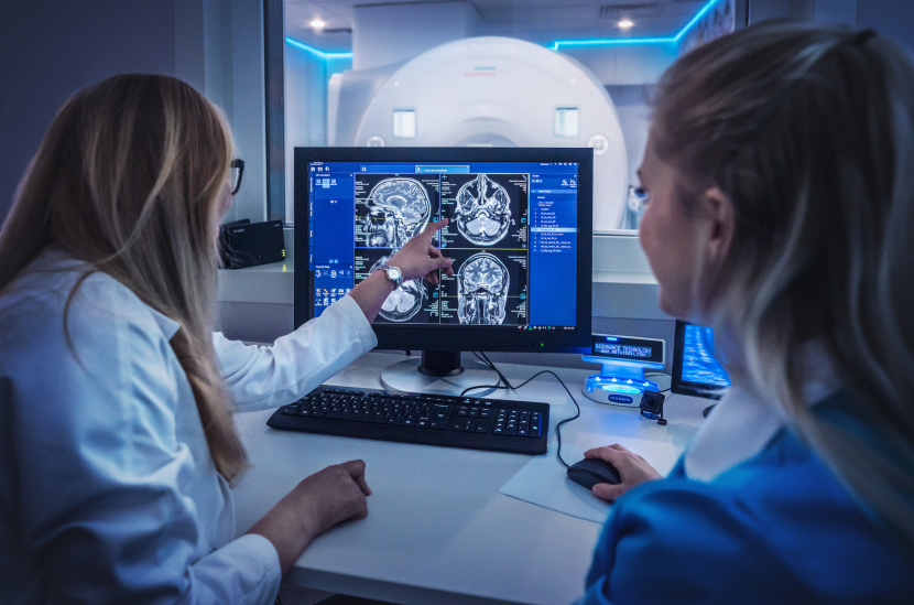
Can you describe the principle of CT and MRI?
Computed tomography (CT) works on the principle of detecting X-rays that pass through the object of interest, in this case the patient, while magnetic resonance imaging works on the principle of a strong magnetic field that causes a temporary change in tissue behaviour, which is then detected by different detectors than a CT scan.
What is the biggest difference and why do patients so often confuse the two devices?
The general public confuses them mainly because the two devices look very similar, even identical. Therefore, when potential patients see the tunnel, they cannot distinguish which imaging modality it is. The interior of the two devices is, of course, very different.
In the CT machine, there is a so-called X-ray tube and detectors which then read the X-rays.
The MRI machine contains magnetic coils that produce a strong magnetic field, but here too there are detectors that sense the resulting signal.
In addition to the technology, there is a big difference in the time of the examination. CT works very quickly. A full-body CT scan is within minutes, whereas an MRI would take a similar scan in tens of minutes.
What is each method most commonly used for?
The skeleton, i.e. the bones, is the strong point of CT imaging. Above all, CT is the method of first choice in traumatic events, when a patient arrives at the hospital after a car accident, for example, to rule out fractures. Besides being the fastest method, it will also show everything needed for quick intervention by the attending physicians.
Soft tissues are also well imaged by CT, especially the abdomen and lungs. Magnetic resonance imaging, on the other hand, is a completely marginal imaging method for showing the lungs and is not commonly used for this type of examination. MRI is best for imaging just those tissues that are minimally different from each other. Examples are the individual structures of the brain. CT can also image the brain, but there is significantly poorer resolution of detail.
Does the preparation for each scan differ?
For an MRI, the basic prerequisite for the examination is that the patient has no ferromagnetic metals in the body. With CT, this is not required, and therefore the patient can come for the examination even with an implanted pacemaker. Therefore, no special preparation is required for the MRI unless otherwise specified by the attending physician or radiology assistant, for example, when administering a contrast agent. Preparation for CT is similar in this case.
What is the disadvantage of each imaging modality?
The disadvantage of CT is the radiation load, although it is very small and almost harmless to humans. However, in some indications this can be decisive, for example in young children.
The disadvantage of magnetic resonance imaging is the longer imaging time, which requires the cooperation of the patient, who must be still for the entire examination. For these reasons, it is very common, especially in young children, to encounter some form of anaesthesia or sedation. In our centre we have tried to solve this problem by using special glasses. Patients have them available throughout the examination and can view selected soothing videos or films, which is especially distracting for young children. At the same time, patients who suffer from claustrophobia also make very frequent use of them.
Is there a diagnosis that you encounter most often?
The most common MRI scans are of the central nervous system, the brain and imaging of the soft tissues of the musculoskeletal system such as the discs in the spine or ligaments in the knees.
For CT scans, it is definitely cancer patients where tumours are diagnosed or the effect of treatment is monitored.
Is there a difference between the different types of MRIs?
All the machines are usually very similar, which is due to the technical parameters of the design. They differ in the strength of the magnetic field, denoted as T (Tesla). The most commonly used instruments are the 1.5T and 3T instruments, where, in addition to the effect of magnetic field strength, differences in imaging quality can also be based on the software and accessories of the instrument.
There are also instruments without the usual tunnel, so-called open tunnel; they usually have a weaker magnetic field, but do not affect sensitive individuals as claustrophobically and some have the option of standing imaging. On the other hand, instruments stronger than 3T are used for scientific purposes rather than routine diagnostics.
At the Diagnostic Centre we have a Siemens Magnetom Vida MRI with a magnetic field strength of 3 Tesla.