Česky
Česky


How does MRI work?
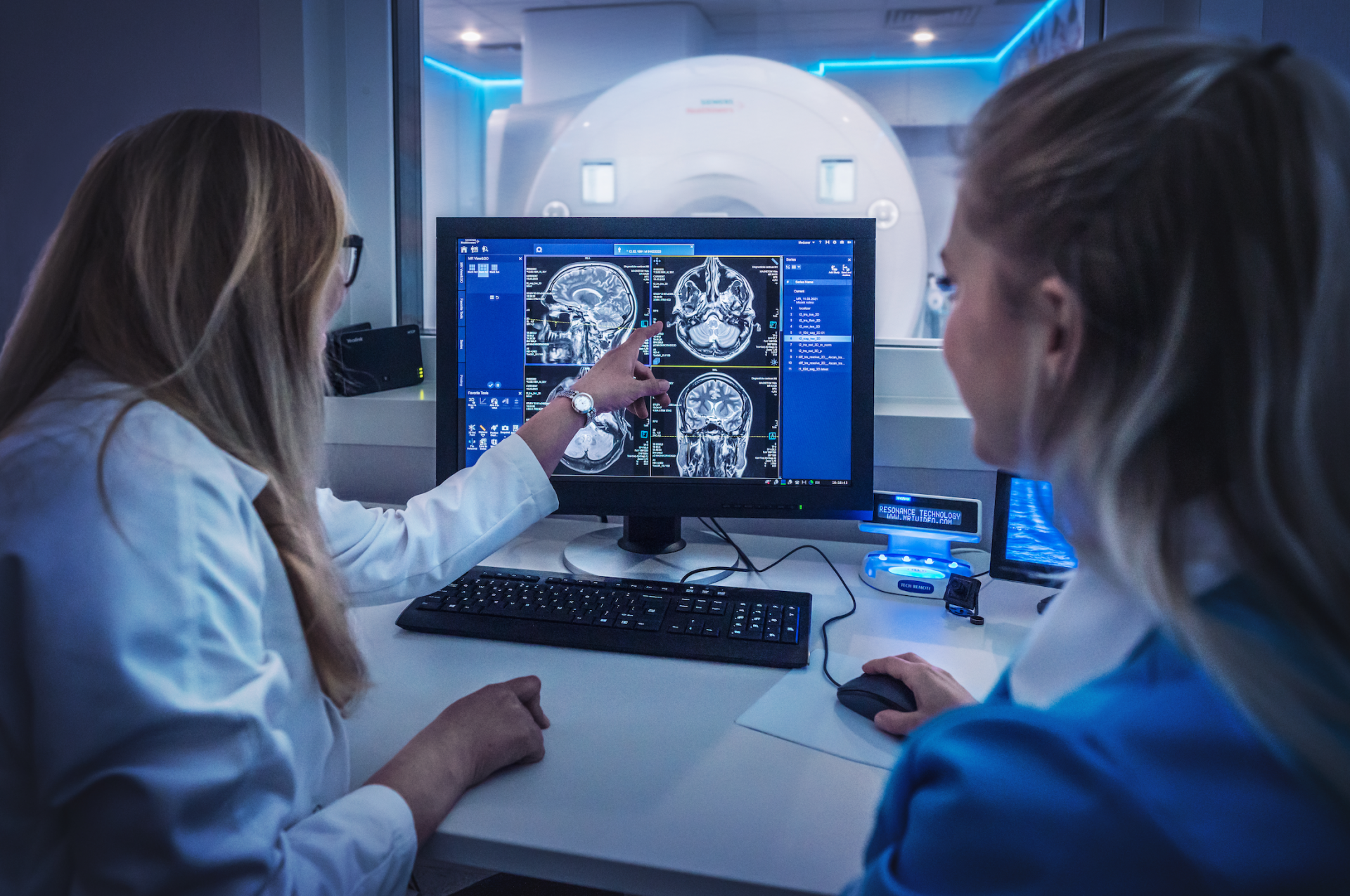
MRI uses a strong magnetic field to image the tissues of the human body. Each part of the human body is made up of different proportions of water, and hydrogen molecules are the element that is most sensitive to magnetic fields. There is a single proton in the hydrogen nucleus that is deflected from its natural rotation by the magnetic field, and then returns to it when the magnetic pulse is turned off. On this return, it sends out high-frequency signals which the instrument picks up and converts the data into a wide range of greyscale images.
What is the difference between MRI and CT?
Unlike MRI, computed tomography (CT) uses ionising radiation to image the human body, with the resulting image depending on the density of the tissues through which the X-rays pass. Computed tomography examinations are generally much faster and are well suited for imaging, for example, bone or lung tissue. However, unlike MRI, the patient is exposed to (albeit very small) doses of ionising radiation, which can have a negative effect on the cells of the human body. The administration of an iodine contrast agent may be necessary to distinguish individual soft tissue structures, with some risk of allergic reactions.
In MRI, contrast agents are also used in indicated cases, but they work on a different principle and allergic reactions are very rare.
Which diseases is it most commonly used for?
Magnetic resonance imaging is indispensable for imaging diseases of the nervous system, especially of the brain or spinal cord. It also provides a detailed diagnosis in musculoskeletal disorders, whether they are prolapsed intervertebral discs of the spine or post-traumatic damage to ligaments of the joints.
However, it is also widely used in oncological diagnostics, for example to assess the extent of cervical, rectal or prostate tumours and to monitor the subsequent effects of treatment.
How is an MRI scan performed?
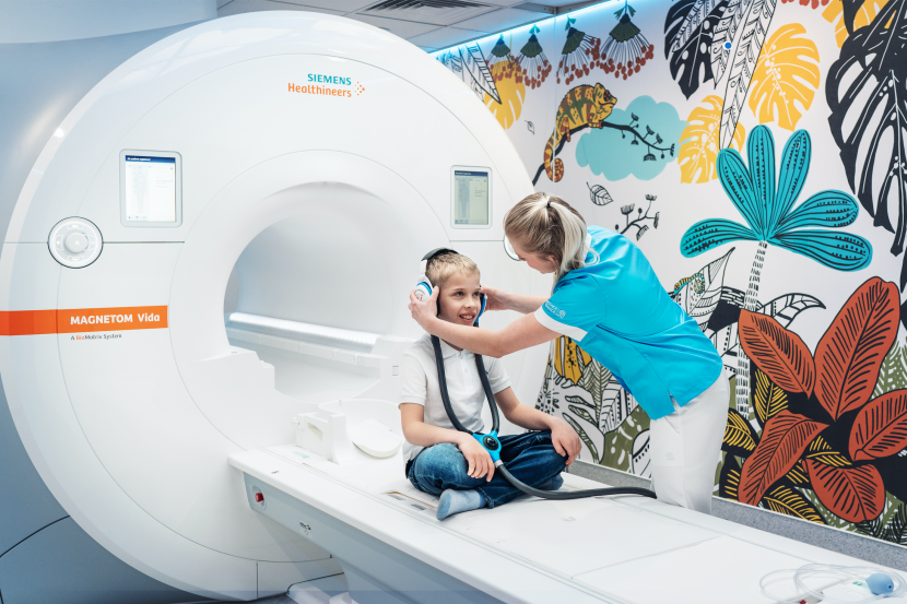
The patient is placed on the movable examination table of the machine, usually on their back. A so-called coil is then attached to the part of the body being examined (e.g. head, knee), which is part of the machine that is necessary to produce a high-quality image. The patient is then inserted into the gantry of the machine (called a tunnel) by a radiology assistant.
The patient's only task is to lie still and not move. The length of the examination depends on the specific indications for imaging, the shortest - around 20 minutes - include, for example, the examination of joints or one section of the spine.
The machine is very loud when running, so patients wear headphones. In addition, patients at our clinic have special glasses, which can be used to project selected soothing videos, such as nature, animals or fairy tales.
Does the patient need any special preparation for the examination?
No preparation is necessary for most MRI examinations. The exception to this is some specialised examinations of the digestive tract, where the patient comes to the examination on an empty stomach.
Who is not suitable for the examination?
The examination is generally contraindicated for patients with pacemakers and cochlear implants. A general contraindication to the examination is if the patient has ferromagnetic metals anywhere in the body or those whose composition cannot be ascertained with valid information.
However, for many years most metals used in surgery or dentistry have been non-magnetic and safe for examination, but it is not advisable to have an MRI scan until six weeks after they have been implanted in the body.
Claustrophobia and an uncooperative patient are relative contraindications, but this can be addressed in selected cases in hospital settings by examination under general anaesthesia.
Are there different types of MRI machines? How do they differ and are some more accurate?
All machines are usually very similar, which is due to the technical design parameters. They differ in the strength of the magnetic field, denoted as T (Tesla). The most commonly used instruments are the 1.5T and 3T instruments, where, in addition to the effect of magnetic field strength, differences in imaging quality can be based on the software and accessories of the instrument.
There are also instruments without the usual tunnel, so-called open tunnel; they usually have a weaker magnetic field, but do not affect sensitive individuals as claustrophobically and some have the option of dynamic standing imaging. On the other hand, instruments stronger than 3T are used for scientific purposes rather than routine diagnostics.
What is the average waiting time for an MRI scan? Why is the wait so long?
The average waiting time for an MRI scan certainly varies from region to region or city to city. In Prague it is often around three months, but it depends on the medical facility. However, the specific indication for the examination is always decisive; i.e. a patient after an accident with a suspected spinal cord injury is usually examined immediately, while diagnoses of long-standing headaches or back pain unfortunately wait longer.
Source: Petra Barochová, iDNES.cz, 4 June 2021